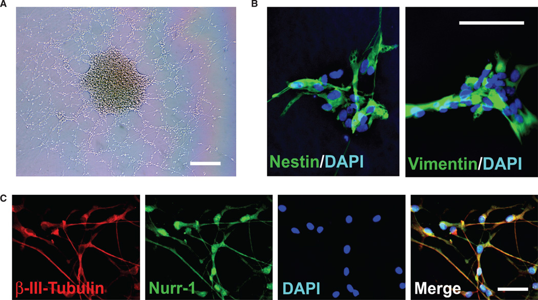
Figure 1.
Primary hNSC isolated directly from the human fetal neuroectoderm. (A) A phase image of the CNS-derived hNSC in culture. (B) The hNSC (>95%) express neural stem/progenitor marker nestin (green) and vimentin (green). All cells are revealed by DAPI staining of their nuclei (blue). (C) When provided with appropriate cues, the hNSC (c. 5–10%) differentiate into neurons [labeled for β-III-tubulin (red)] that express Nurr-1 (green), a marker associated with the midbrain dopaminergic phenotype. Scale bars: (A) 50 µm; (B, C) 5 µm.

