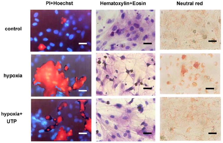
Fig. 4.

Effect of UTP on cardiomyocyte morphology in hypoxic conditions. Six-day-old cultured cardiomyocytes under normoxic conditions were subjected to 2 h hypoxic environment or pretreated with 50 μM UTP and then subjected to hypoxia. One group of these cells was stained with propidium iodide (red), which marks damaged cells, and with Hoechst 33342 (blue), which stains live-cell nuclei (first column). A second group of cells was stained with hematoxylin and eosin (second column). A third group of cells was stained with neutral red, which stains lysosomes (third column). The results shown are representative of six experiments. Bars = 10 μm.
