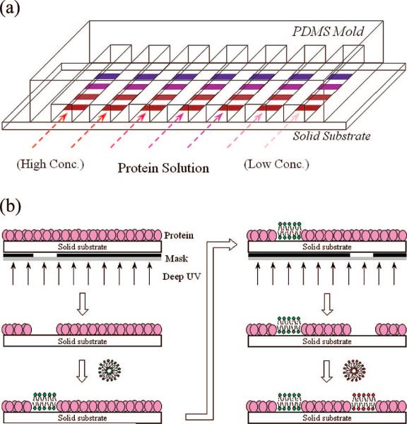
Figure 1.
(a) Schematic representation of a seven-channel microfluidic device. Each channel is addressed with four distinct lipid bilayers designated by colored patches. Aqueous solutions with various protein concentrations are arrayed from left to right. (b) Schematic diagram of the sequential patterning technique employed for creating spatially addressed bilayer arrays.

