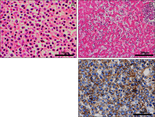
Figure 2 .
Histological and immunohistochemical examination of DLBL. (A) On high-power view, these tumor lesions were composed of a diffuse and monotonous proliferation of large to medium-sized atypical lymphoid cells having enlarged hyperchromatic nuclei and prominent nucleoli. (H&E stains, Original magnification × 400, Bar = 50 μm) (B) In the liver, not only nodular growth pattern but sinusoidal proliferation of the lymphoma cells were evident. (H&E stains, Original magnification × 100, Bar = 200 μm) (C) Immunohistochemically, these atypical cells were diffusely positive for CD20. (Original magnification × 400, Bar = 50 μm).

