
Figure 5. Src inhibition with dasatinib effectively inhibits tumor growth in vivo.
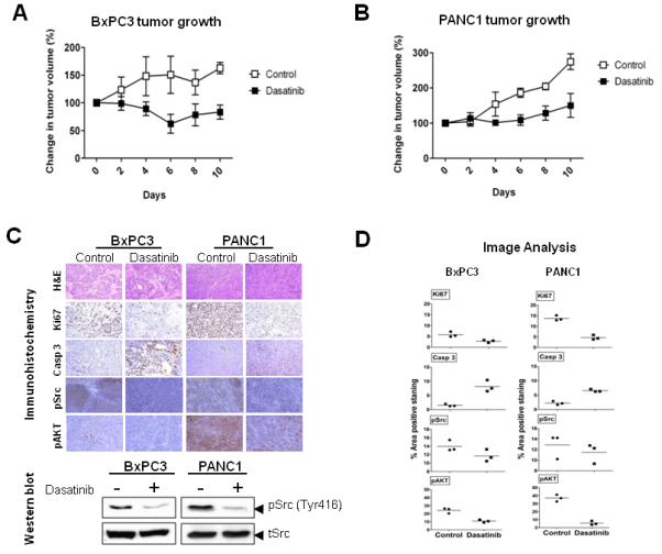
Mice with subcutaneously established tumors from BxPC3 (A) or PANC1 (B) cells were treated with dasatinib (n = 5) at 25 mg/kg or vehicle (n = 5) daily by oral gavage for 10 days. Growth curves for tumors are presented as the mean ± SD of five tumors in each data point. (C) Representative examples of immunohistochemical analysis of BxPC3 and PANC1 tumor tissues stained with H&E, Ki67, cleaved caspase 3, pSrc and pAKT. There is no evidence of treatment-induced necrosis on H&E staining. Compared with vehicle treated controls, Ki-67, pSrc and pAKT staining is markedly decreased, and cleaved caspase 3 staining is markedly elevated in dasatinib treated animals, showing successful target inhibition by dasatinib. Magnification ×20. Tumor tissue was analyzed for the expression of pSrc by Western blotting. (D) The percent area positive staining was determined using Image J image analysis software for Ki67, cleaved caspase 3, Phospho Src (pSrc) and phospho AKT (pAKT) stained immunohistochemistry data from BxPC3 and PANC1 tumor xenografts. Individual data points represent the mean ± SD of three independent tissue samples analyzed in each treatment.