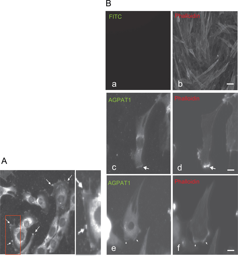
Fig. 2.
Subcellular localization of AGPAT1 in myoblast. (A) Immunostaining of endogenous AGPAT1 in C2C12 showing AGPAT1 accumulation at the sites of membrane extensions (arrows); magnification 60×. (B) Immunostaining for AGPAT1 and phalloidin in C2C12 showing the accumulation of AGPAT1 and actin filaments at the leading edge (arrows). (a,b) Immunostaining only for phalloidin. Bar: 50 uM. (c–f) Immunostaining for phalloidin and AGPAT1. Bar: 10 uM.

