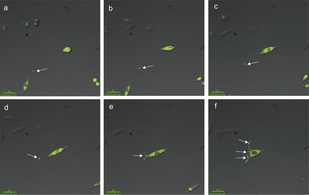
Fig. 4.
Live time-lapse imaging reveals that AGPAT1 accumulates at the distal tips of filopodia in C2C12 myoblasts. C2C12 cells expressing GFP tagged AGPAT1 were imaged every 10 min for 16 h. AGPAT1 concentrated at the distal tips of filopodia (arrow). There was also accumulation at areas of contact with other myoblast (e,f). Bar: 40 uM.

