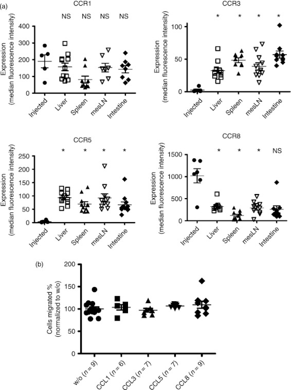
Figure 2.

Expression of chemokine receptors on regulatory T (Treg) cells isolated from different tissues. (a) Surface expression of the indicated receptors, after gating on CD4+ FoxP3+ cells is displayed for freshly isolated Treg cells from spleen of naive animals or cells isolated from the organs liver, spleen, mesenteric lymph nodes or intestines of transplanted animals. The Treg cells that were isolated from naive donor mice were termed ‘injected’, the Treg cells isolated from the different organs were named according to the organ they were isolated from. Each data-point represents an individual donor mouse (n = 9; *P < 0·0001). The experiment was performed twice and the results were pooled. (b) In vitro migration of Treg cells was assessed in a transwell chamber with or without CCL1, CCL3, CCL5 and CCL8 as a stimulus (*P < 0·05). The experiment was performed three times with similar results.
