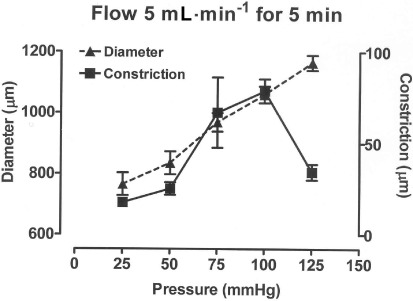
Figure 3.

Graph showing how the external diameter and the constriction induced by flow at 5 mL·min−1 for 5 min varied over a range of distending pressures (25–125 mmHg) in carotid artery segments. All vessels were initially set at a low level of U46619-induced tone while at a pressure of 100 mmHg. Data are presented at the mean ± SEM of 5 observations.
