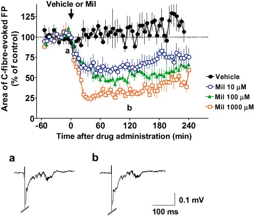
Figure 4.

Effects of spinally administered milnacipran (Mil) on the basal C-fibre-evoked field potentials (FPs) in modified spinal nerve ligation (SNL)-model animals. FPs in the spinal dorsal horn were elicited by electrical stimulation of the sciatic nerve fibres at 1 min intervals in modified SNL-model animals. The vehicle (n= 5) or 10, 100 and 1000 µM milnacipran (n= 5, n= 7 and n= 4, respectively) was administered spinally after ≥60 min of stable baseline recordings (arrow). Each area of C-fibre-evoked FPs was normalized to the mean of 60 consecutive responses obtained prior to the administration (–60 to 0 min in the graph) and five consecutive responses were averaged. Example-averaged traces of five consecutive FPs from the 10 µM milnacipran group recorded during the times indicated on the graph (a and b) are shown. Data shown are means ± SEM.
