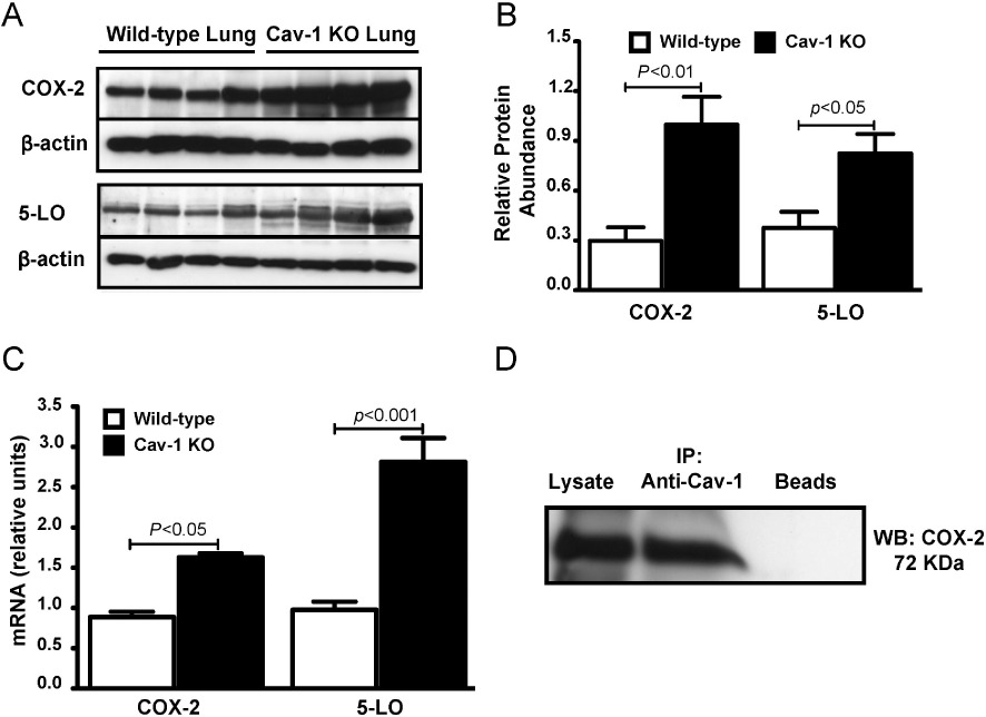
Figure 4.

Increased expression of COX-2 and 5-LO in lungs from Cav-1 KO mice. (A) Representative Western blots and (B) corresponding densitometric analyses showing COX-2 and 5-LO abundance in whole lung lysates from Cav-1 KO and wild-type mice (n- 4–5 for each group). P-values shown were obtained after one-way anova with Tukey's multiple comparison test. (C) Histogram showing results of quantitative RT-PCR for COX-2 and 5-LO mRNA in lysates from lungs of Cav-1 KO and wild-type mice. 18S RNA was used as an internal control (n- 4–5 for each group). P-values shown were obtained after one-way anova with Tukey's multiple comparison test. (D) Western blot (WB) showing COX-2 abundance after immunoprecipitation (IP) with Cav-1 antibody and protein-G-conjugated sepharose beads from lung homogenates of wild-type mice. The lane labelled ‘Beads’ included sample but no antibody.
