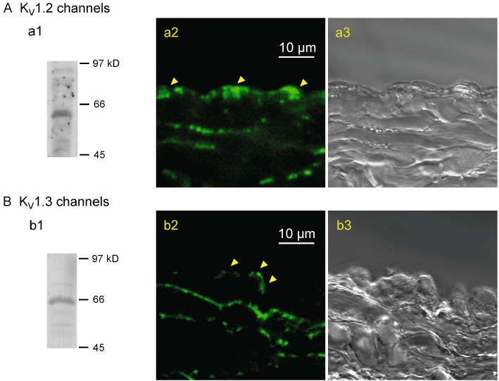
Figure 10.

Expressions of KV1.2 and KV1.3 channels in rabbit jugular vein. (A) KV1.2, (B) KV1.3. Western blots to show the specificities of the antibodies against KV1.2 (∼60 kDa) (a1) and KV1.3 (∼65 kDa) (b1) channel proteins. Fluorescence images showing localization of antibodies against either the KV1.2 (a2) or KV1.3 channels (b2) in cross-sections of jugular vein, together with the corresponding bright-field images (a3 and b3). Arrows indicate fluorescence signals in endothelial cells. Similar observations were made in three other preparations.
