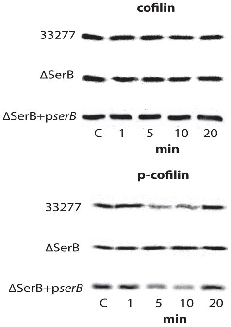
Figure 1. Western blot analysis of cofilin and p-cofilin levels in P. gingivalis–infected HIGK cells.
Whole cell lysates of P. gingivalis 33277-infected, P. gingivalis ΔSerB-infected, or P. gingivalis ΔSerB+pserB-infected HIGK cells were examined by Western blotting with antibodies to cofilin (upper panel) or phospho(p)-cofilin (lower panel). Data are representative of three independent experiments.

