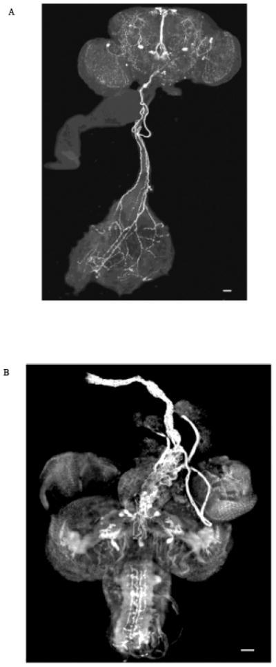
Fig. 4.

Staining of (A) adult CNS and gut and (B) pupal CNS and anterior dorsal vessel (aorta) identify immunoreactive material as fluorescent signal (white). DMS-specific antisera stained neurons in the (A) subesophageal ganglion and (B) protocerebrum which sent immunoreactive fibers to innervate the gut and aorta, respectively. The bars in the lower right-hand corner represent 50 μM.
