Fig. 4.

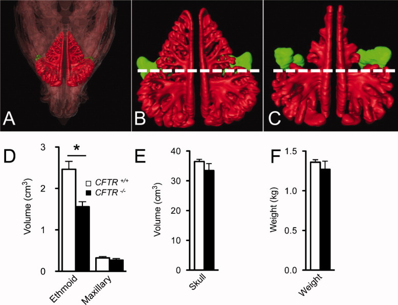
Cystic fibrosis (CF) piglets have ethmoid hypoplasia at birth. (A) Three-dimensional reconstruction view of skull (transparent overlay) and sinus of newborn pig. (B) Non-CF and (C) CFTR−/− newborn piglet three-dimensional sinus scan with ethmoid (red) and maxillary (green) sinus. Dotted line dividing anterior and posterior ethmoid sinus. Volumetric analysis performed on three-dimensional reconstructions of segmented sinus computed tomography scans. Data points represent mean ± standard error of non-CF (n = 7; open symbols) and CFTR−/− (n = 8; closed symbols) littermate pigs. *P < .05. (C) Volume of ethmoid and maxillary sinuses. (D) Volume of skull. (E) Weight of newborn pigs.
