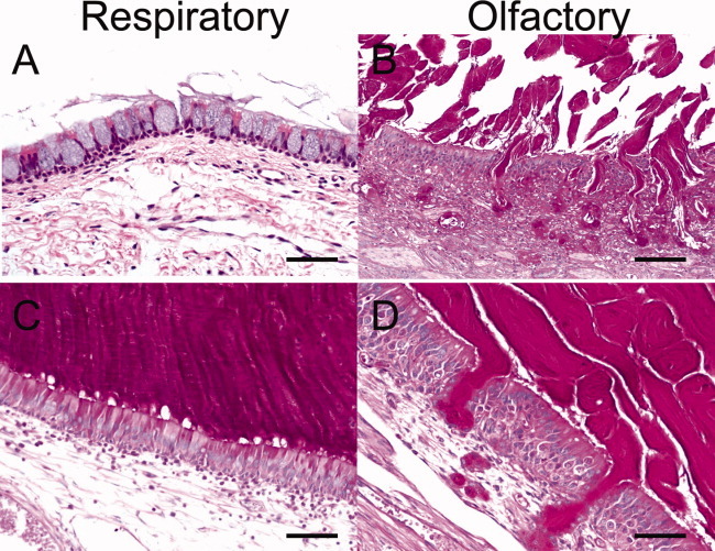
Fig. 7.

Cystic fibrosis (CF) pig sinus disease shows mucus accumulation and epithelial remodeling on histology. (A, B) Moderate sinus disease in CF pigs showed goblet cell hyperplasia with mucus accumulation and submucosal gland hypertrophy, periodic acid-Schiff (PAS) stain, scale: 50 μm. (C, D) Severe sinus disease with mucus from goblet cells and submucosal glands, PAS stain, bar: 55 μm.
