
Fig. 2.
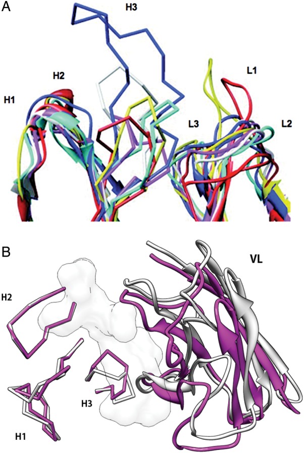
Structural diversity of antibodies. (A) Variability of six CDRs. (B) Structural changes of CDR-H3 [root mean square deviation (RMSD) of backbone N, Cα, C and O atoms is 2.3 Å] and rearrangement of VL/VH domain orientation. Anti-HIV-1 peptide antibody, Fab50.1 (magenta (1GGI) and white (1GGC) for antigen bound and free structures, respectively). The white surface model is the antigen, HIV-1 V3 loop peptide. All graphics of protein structures in this article are generated using UCSF Chimera (http://www.cgl.ucsf.edu/chimera).