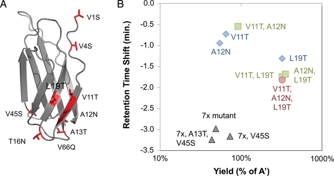
Fig. 3.
Multiple mutant analysis. (A) The NMR structure of the tenth type III domain of human fibronectin (1TTG (Main et al., 1992)) is presented in cartoon form with sites of beneficial mutation depicted in red. (B) Mutant clones were produced in BL21(DE3), purified, and analyzed by RP-HPLC using a 10–90% acetonitrile gradient over 30 min. The yield and retention time shift, both relative to the parental clone, are indicated for single (blue diamonds), double (green squares) and triple (red circle) mutants of V11T, A12N and L19T. The 7x mutant (V1S, V4S, V11T, A12N, T16N, L19T, V66Q) and derivatives are shown in black triangles.

