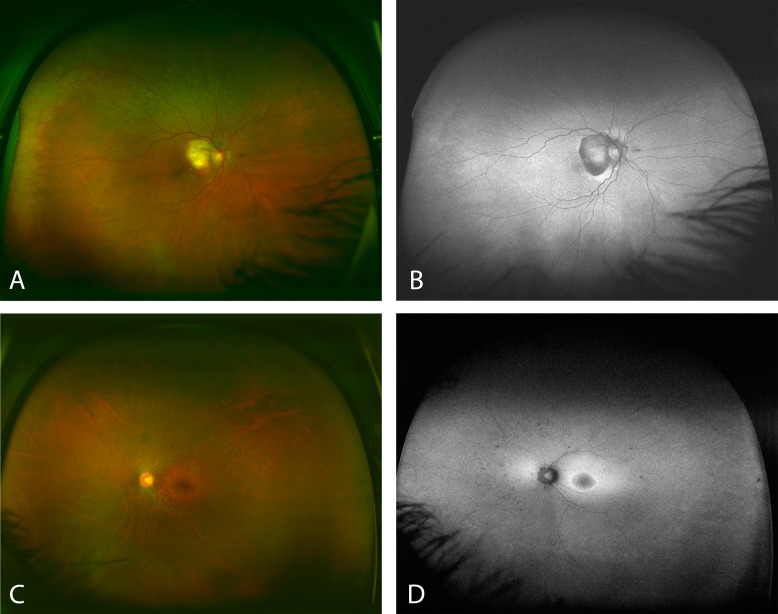
Figure 2. .
Images (A) and (B) show a case with AMD that appears to have almost no peripheral abnormalities on FAF (B), but the abnormalities seem more evident in the pseudocolor image (A). Contrarily, (C) and (D) show a case with a retinal dystrophy. The pseudocolor image (C) does not suggest noticeable peripheral abnormalities. However, they become more clear on the FAF image (D).

