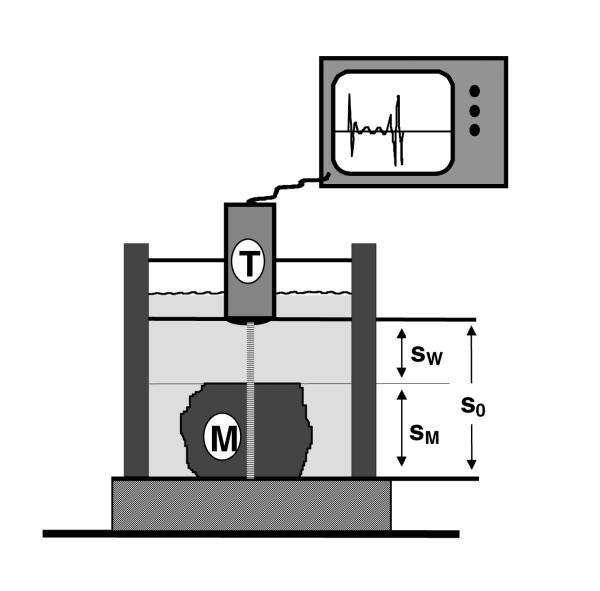
Figure 1.
Schematic representation of the sonographic device. A 20-MHz transducer (T) is fixed to a basin filled with water. The 12-millimeter-distance (so) between the transducer and the plexiglas bottom can be measured with or without interposing tissue samples. The melanoma tissue (M) can be fixed onto the bottom with a flexible clip in different positions.

