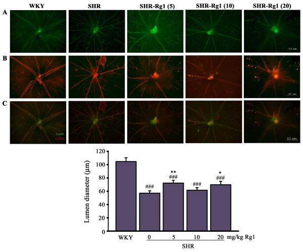
Figure 3.
Effects of Rg1 on retinal remodeling induced by hypertension. Whole-mount double-staining of retinal vessels with FITC-coupled lectin (green) and α-SMA antibody (red). (A) lectin positive arteries. (B) α-SMA–positive arteries. (C) The merged picture for both (A) and (B). (D) Quantitative data of lumen diameter for retinal arteries. ###p < 0.001 compared with WKY; **p < 0.01, **p < 0.01 compared with SHR. n = 4 for each group.

