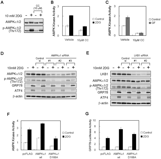
Figure 4. Effects of the AMPK kinase activity inhibition on GRP78 accumulation.
(A) Immunoblot analysis. HT1080 cells were treated with compound C for 4 h in the presence (+) or absence (−) of 10 mM 2DG. β-actin was used as a loading control. (B, C) AMPK kinase assay. HT1080 cells were treated with compound C for 2 h under 2DG stress (B) or glucose withdrawal (C) conditions. CC, compound C; GF, glucose-free. Results shown are the means ± SD of triplicate determinations. (D, E) Immunoblot analysis. HT1080 cells were transfected with AMPKα1 (D) or LKB1 siRNA (E) and cultured for 18 h in the presence (+) or absence (−) of 10 mM 2DG. β-actin was used as a loading control. (F) AMPK kinase assay. HT1080 cells were transfected with plasmid subcloned wild-type AMPKα1 or dominant negative D168A, and cultured for 2 h under normal or 2DG stress conditions. A pcFLAG vector was used as a control. Results shown are the means ± SD of duplicate determinations. (G) Reporter assay. HT1080 cells were co-transfected with pGRP78pro160-Luc and plasmid-expressed wild-type AMPKα1 or dominant negative D168A and were cultured for 18 h under normal or 2DG stress conditions. A pcFLAG vector was used as a control. Relative ratios of 2DG-induced promoter activities were calculated by setting normal activation level in each cell as 1. Results shown are the means ± SD of quadruplicate determinations. 2DG, 10 mM 2-deoxy-D-glucose.

