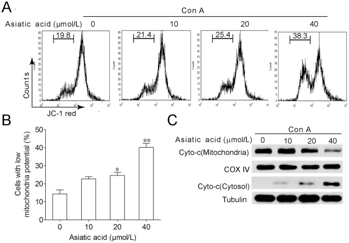
Figure 3. Asiatic acid triggers apoptosis of Con A-activated T cells through mitochondrial pathway.
Lymph node-derived T cells isolated from BALB/c mice were incubated in medium or in the presence of Con A (5 µg/ml) for 24 h. Then cells were further incubated with or without various concentrations of asiatic acid for 24 h. Cells were stained with JC-1 intensity of FL-2 was analyzed by flow cytometry to determine mitochondrial membrane potential. The cells located with low red fluorescence were considered as low mitochondrial transmembrane potential (A). Data are shown as means ± SEM of three independent experiments (B). *P<0.05, **P<0.01 vs. drug-untreated group. (C) The release level of cytochrome c from mitochondria was examined by Western blotting. The results shown are representative of three experiments.

