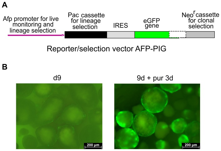
Figure 1. Differentiation and pre-selection of eGFP-expressing cells in a rotary EB culture.
(A) Essential part of the AFP-PIG vector (“PIG”: after Pac-IRES-eGFP). (B) eGFP-positive cells in the outer rim of 9-day-old EBs (left panel) and appearance of expanding clusters of eGFP-expressing cells after a 3-day treatment with puromycin (right panel).

