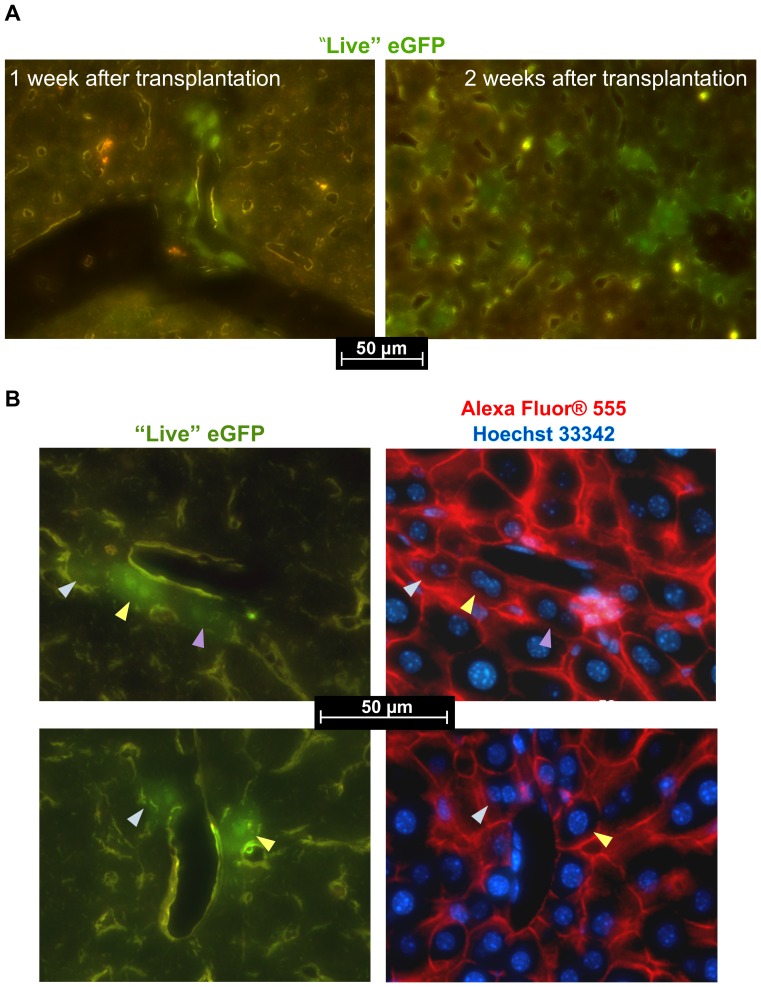
Figure 6. Engrafted spheroid-derived cells within recipient liver tissue.
(A) eGFP-fluorescent cells and cell clusters observed in liver sections one and two weeks after spheroid transplantation. (B) Expression of Ecad in engrafted cells. “Live” eGFP and merged Alexa Fluor® 555/Hoechst 33342 images are shown.

