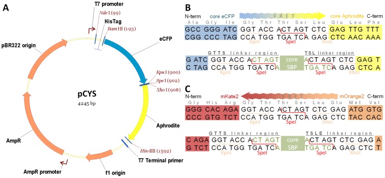
Figure 1. FRET vector and insertion sites for cloning SBPs.
(A) vector pCYS and cloning sites of (B) pCYS and (C) pROS. Direction of energy transfer between fluorescent proteins is shown. Fluorescent proteins are coloured coded; N-terminal His-tagged-eCFP (blue), Aphrodite (yellow), N-terminal His-tagged-mKate2 (red), and mOrange2 (orange). The position of core SBP insertion (green) is shown, together with the amino acid sequences of N-terminal (GTTS) and C-terminal (TSL/TSLE) linkers.

