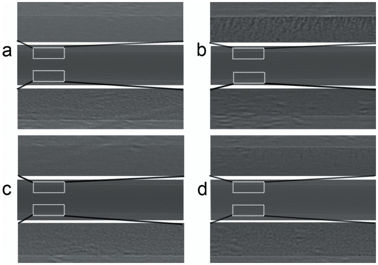
Figure 7. PCI of LLC cells isolation with MBV (a–b) or MBC (c–d).
(a and c) Before introducing the magnetic field, the mixture of cells and microbeads were clearly revealed along the lower edge of the PE-50 tube. (b and d) After placing a magnet over the tube, the bead-bound cells were attracted towards the upper edge of the tube. Note that the MBV-bound cells (b) were much more than MBC-bound ones (d). Images were obtained at the energy of 14 keV. The pixel size was 0.74 µm ×0.74 µm.

