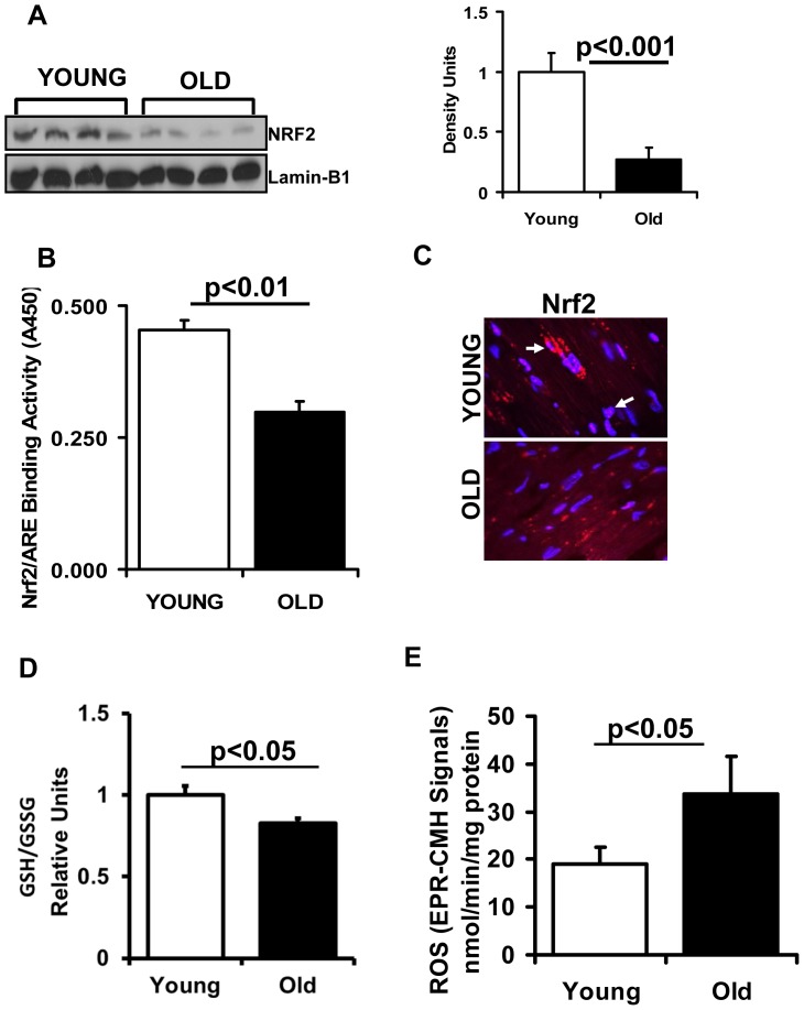
Figure 1. Decreased function of Nrf2 in the aging heart.
(A) Decreased nuclear translocation of Nrf2 is evident in the heart of aging mice. Representative immunoblot (IB) of nuclear proteins collected from young and aged WT mice (n = 6) under basal state. Each lane represents an individual animal/heart. Lamin-B1 used as loading control. Densitometry analysis of the IB images was achieved using Image-J. (B) Transcription factor binding (TransAM-Nrf2 activity) assay: Nuclear proteins from young and aged mice (n = 4) were incubated with the oligonucleotide (pre-coated on 96 well plate/strips) for ARE (antioxidant response element). (C) Localization of Nrf2 by immunofluorescence: Immunofluorescence analysis using anti-Nrf2-ab (1∶200; v/v) showing decreased cytosolic and nuclear Nrf2 in old versus young myocardium. Blue: nucleus (DAPI); Red: Nrf2-staining and Pink: merge of blue and red indicating nuclear localization of Nrf2. IF images were obtained at a magnification of 60X oil immersion. (D) Glutathione redox-state: Myocardial redox state was determined in the ventricles of 2 and 23 months old mice under basal conditions. Statistically significant changes in the redox ratio (GSH/GSSG) were observed in young and old groups. Values are mean ± SD for 5 or more animals in each group. (E) Increased reactive oxygen species (ROS) generation in the hearts of aging mice: Electron paramagnetic resonance (EPR) spectroscopy signals for CMH (1-hydroxy-3-methoxy-carbonyl-2, 2,5,5- tetramethyl pyrrolidine) in young and aged mice. EPR signals for CMH are significantly increased in aged versus young mouse myocardium. Values represent n = 5 or more from each group.

