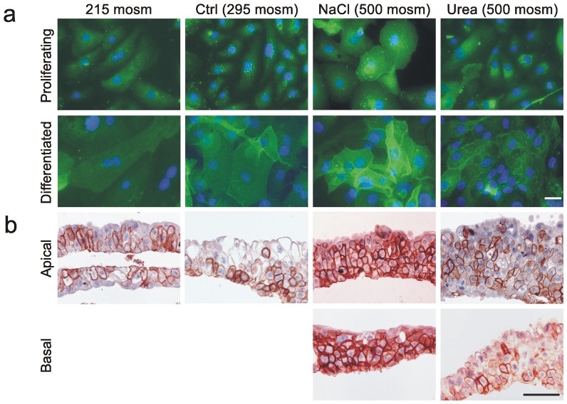
Figure 1. Immunolocalisation and expression of AQP3 in response to altered medium osmolality.
a) Immunofluorescence labelling of non-differentiated (proliferative) and differentiated NHU cell cultures grown on glass slides showing increased labelling intensity and membrane localisation of AQP3 at high concentrations of NaCl, but not urea, following 72 hours of exposure. Scale bar: 10 µM. b) Immunohistochemistry of differentiated NHU cell constructs grown on permeable Snapwell membranes and exposed for 72 hours to indicated osmolalities. Note no effect of urea, but major increase in AQP3 expression in all layers in response to medium made hyperosmotic (500 mosm/kg) by the addition of NaCl, irrespective of exposure via apical or basal aspect. Scale bar: 50 µM.

