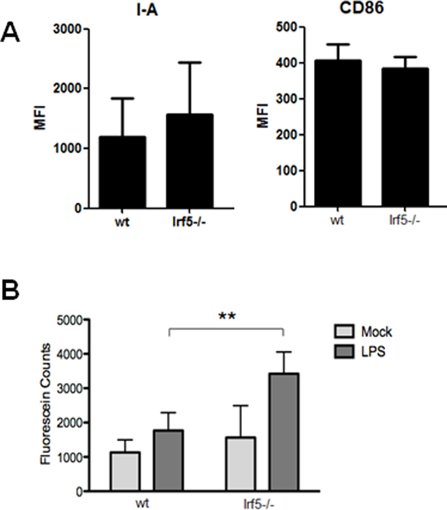
Figure 6. CD11b+ splenic macrophages from Irf5−/− mice display enhanced phagocytosis.

(A) PECs were collected from Irf5+/+ (wt) and Irf5−/− littermates 4 wks post-pristane injection. Expression of activation/maturation surface markers I-A (MHC II) and CD86 were examined on CD11b+Ly6G− monocytes. n = 5 mice per genotype. (B) Splenic macrophages from Irf5+/+ and Irf5−/− mice were isolated by positive selection and in vitro phagocytosis determined by measuring fluorescein counts. n = 4–5 per genotype; **p < 0.01 by unpaired Students t test.
