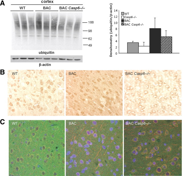
Figure 10.
Ubiquitination levels increase in BACHD and BACHD Casp6−/− mice. A, Western analysis shows an increase in overall protein ubiquitination levels in 13-month-old cortex (left panel). Densitometry of total ubiquitinated protein normalized to β-actin (right panel; n = 3). B, Immunohistochemistry shows changes in localization of ubiquitinated proteins in 15-month-old BACHD Casp6−/− cortex. C, Double immunofluorescence of ubiquitin (red) and huntingtin (green) confirms colocalization in 13-month-old cortex. DAPI nuclear stain is in blue. Error bars indicate SD.

