
Figure 9.
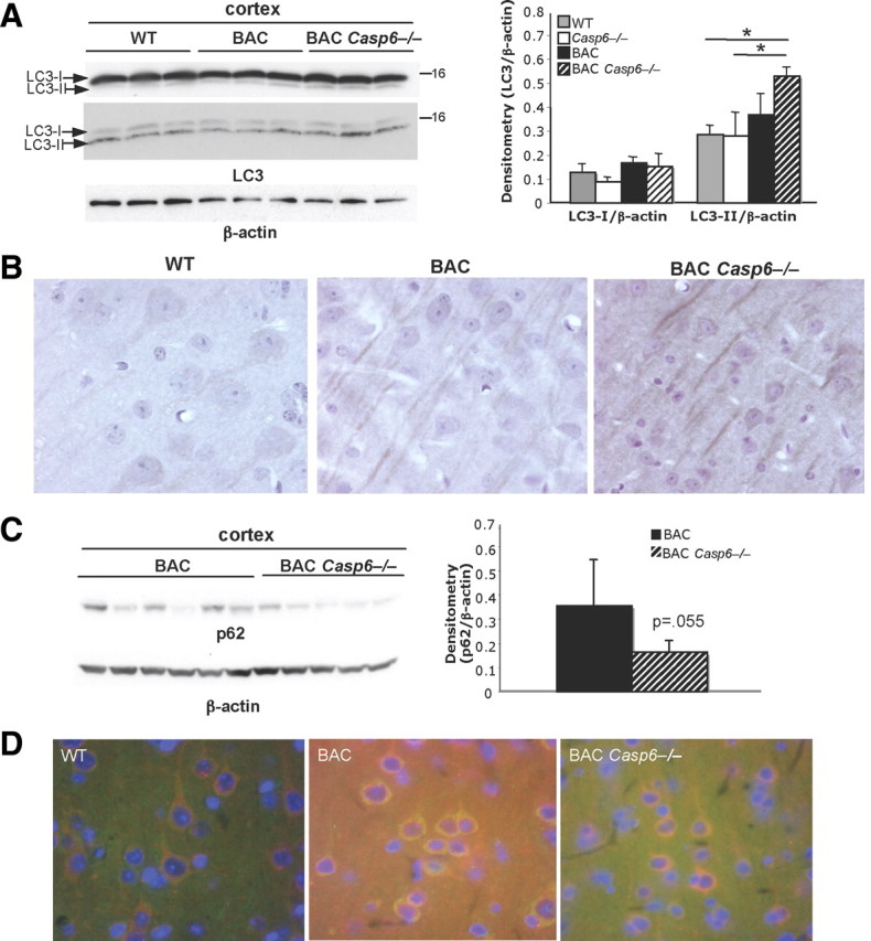
Autophagy-associated proteins are altered in 13-month-old BACHD Casp6−/− mice. A, Western analysis shows an increase in LC3-II levels in BACHD Casp6−/− cortex with antibodies from MBL (top left panel) and Abgent (middle left panel). Densitometry of cortical LC3 levels (Abgent antibody) normalized to β-actin (right panel; n = 3; ANOVA, *p < 0.05). B, Immunohistochemistry shows an increase in LC3 immunoreactivity and altered localization in the 13-month-old BACHD Casp6−/− cortex relative to both WT and BACHD. C, p62 protein levels decrease in BACHD Casp6−/− cortex relative to BACHD (left panel; 13- and 15-month-old cortex) using Western blot analysis. Densitometry of p62 normalized to β-actin (right panel; n = 5–6; t test). D, Immunohistochemistry shows colocalization of p62 (red) and Htt acetylated at amino acid K444 (green) in 13-month-old cortex. Error bars indicate SD.