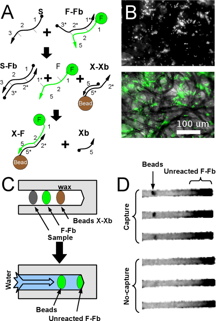
Figure 3. Signal immobilization in the paperfluidic device.
(A) Schematic of the reaction. Released fluorescent oligonucleotides can be captured at specific sites within the paperfluidic device, via beads containing antisense oligonucleotides. (B) Micrographs of immobilized beads. Beads caught in the paper fibers (fluorescence micrograph, top, brightfield, bottom) capture fluorescent oligonucleotide. (C) Schematic of paperfluidic device for detection by oligonucleotide capture on beads. A detection region (Beads X-Xb) is embedded into the paper just downstream of the DNA reagents. (D) Fluorescence image of successful capture showing residual, unreacted F-Fb downstream of bead spots.

