Figure 7.
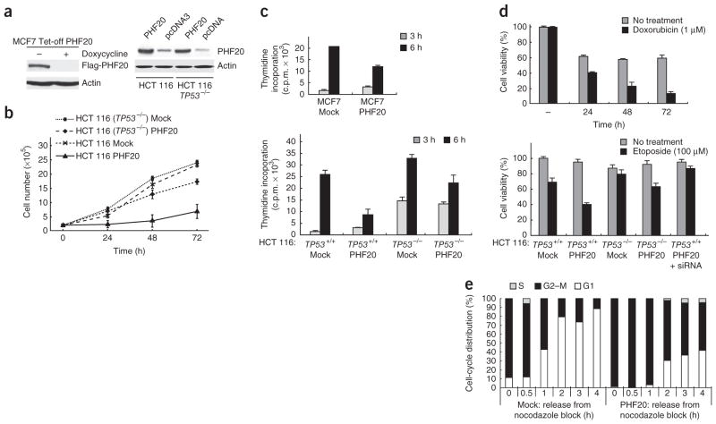
Ectopic expression of PHF20 leads to phenotypic changes consistent with p53 activation. (a) Western blot (WB) analysis showing controlled expression of Flag-PHF20 in MCF7 Tet-off cells (MCF7 Tet-off PHF20). HCT 116 (TP53+/+) and HCT 116 (TP53−/−) cells were transfected with a plasmid encoding PHF20 or empty plasmid pcDNA3, and proteins in the cell lysates were identified by WB using anti-PHF20 and anti-actin antibodies (right). (b) HCT 116 (TP53+/+) and HCT 116 (TP53−/−) cells transfected with a PHF20-encoding plasmid or pcDNA3 (Mock) were grown and counted at the indicated times. Error bars, s.d. from triplicate experiments. (c) Incorporation of 3H-enriched thymidine in MCF7, HCT 116 (TP53+/+) and HCT 116 (TP53−/−) cells transfected with a PHF20-encoding plasmid or pcDNA3 (Mock) was monitored at the indicated times. Error bars, s.d. from triplicate experiments. (d) PHF20-encoding-plasmid–transfected or pcDNA3-transfected (Mock) MCF7 Tet-off, HCT 116 (TP53+/+) or HCT 116 (TP53−/−) cells were treated with 1 μM doxorubicin for the indicated times or with 100 μM etoposide for 4 h. Error bars, s.d. from triplicate experiments. (e) Flow cytometry analysis of fixed MCF7 Tet-off PHF20 cells and control MCF7 Tet-off cells (Mock) that had been cultured in the absence of doxycycline for 12 h and subsequently treated with nocodazole for 12 h, washed and grown in fresh nocodazole-free medium for 1–4 h.

