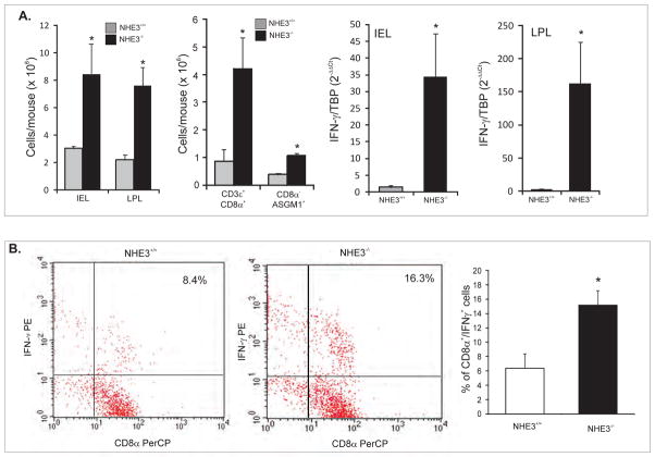
Figure 1. The source(s) of IFN-γ in the small intestinal mucosa of NHE3−/− mice.
(A) Yields of intraepithelial (IEL) and lamina propria (LPL) mononuclear cells, and LPL CD8α+ or CD8α−ASGM1+ cells isolated from the small intestine of WT and NHE3−/− mice, and real-time PCR analysis of IFN-γ mRNA expression in IEL and LPL cells from WT and NHE3−/− mice. (B) Flow cytometry analysis of intracellular IFN-γ in magnetically sorted CD8α+ cells stimulated in vitro with PMA and ionomycin in the presence of brefeldin A. Bar graph depicts summary of four independent experiments.

