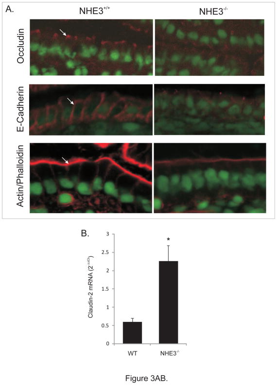
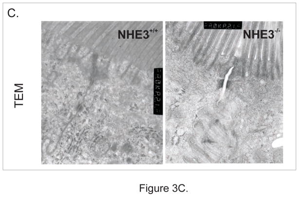
Figure 3.
(A) Immunohistochemical analysis of occludin, E-cadherin, and phalloidin staining of polymerized F-actin in the small intestinal epithelium of WT and NHE3−/−mice. (B) Real-time RT-PCR analysis of claudin-2 expression in the mucosa of WT and NHE3−/− mice. (C) Representative TEM image of the apical junction complex area in the epithelium of WT and NHE3−/− mice.


