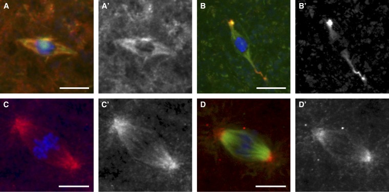
Figure 1 .
KLP10A localization in the female meiotic and embryonic mitotic spindles. Spindles from late-stage oocytes fixed with formaldehyde/heptane (A and B) and syncytial-stage embryos fixed in methanol (C and D) were examined for the localization of endogenous KLP10A (A and C) and HA-tagged KLP10A (B and D). The HA-tagged KLP10A (B and D) was expressed in a wild-type background. (A) In oocytes, endogenous KLP10A localized throughout the meiotic spindle. (B) HA-tagged KLP10A localized throughout the meiotic spindle, but was heavily concentrated at spindle poles. In addition, the “curly pole” phenotype caused by expression of the transgene is observable (see Figure S1). (C and D) In embryos, KLP10A primarily concentrates toward the spindle poles. Microtubules were not imaged in C. In all images, DNA is shown in blue and microtubules are shown in green. KLP10A is in red in merged images (A–D) and in white in single channel images (A′–D′). Bars, 5 μm.

