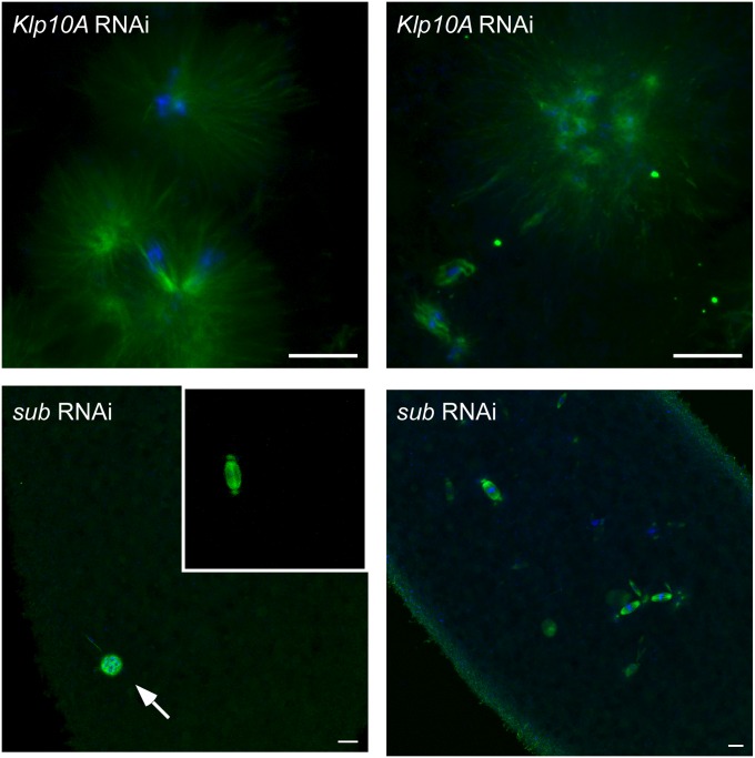
Figure 3 .
Microtubule and DNA disorganization in Klp10A germline mutant embryos. Embryos produced by Klp10A germline mutants show severely disorganized DNA and microtubule structures. Chromosomes are dispersed throughout the cytoplasm, and microtubules form large asters surrounding the dispersed chromosomes. See Figure 1 for wild-type embryo spindles. Also shown are two examples of embryos lacking Subito (by RNAi, see Materials and Methods). About half of the embryos show only the female polar body (arrow) and the male pronucleus (inset). Drosophila female meiosis does not segregate chromosomes into a separate polar body. In the other half of the embryos, there are nuclei attempting to divide, which may have originated from the haploid male genome. DNA is in blue and microtubules are in green. Bars, 10 μm.

