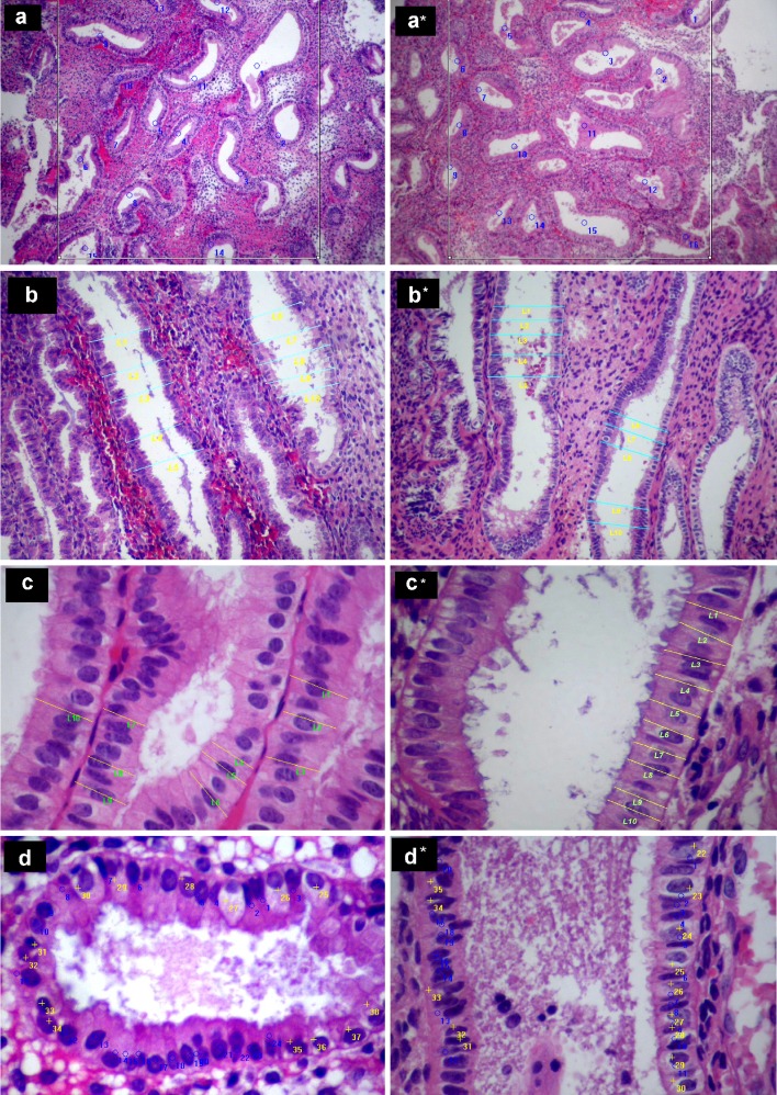
Fig. 1.
Histological appearance, morphometric analysis in endometrial specimens from natural (a, b, c and d) and GnRH-antagonist treated (a*, b*, c* and d*) cycles. a, a* The number of endometrial glands per square millimeter. b, b* The maximal cross-sectional diameter of endometrial glands which were cut parallel to their longitudinal axis. c, c* The height of the glandular epithelial cells. d, d* The number of vacuolated cells (yellow marks) per 1,000 glandular cells

