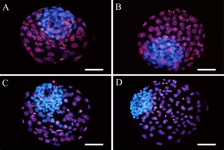
Fig. 2.
Differential stained inner cell mass and trophectoderm cells of bovine day 8 blastocysts developed from washed light mineral oil overlay (A), WMO overlay plus co-culture (B), sterile filtered light paraffin oil overlay (C) or SPO overlay plus co-culture (D). All embryos were healthy with numerous cell numbers. Scale bars: 100 μm.

