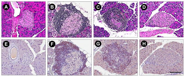
Figure 5.
Photomicrograph of representative H&E (top) and CD8+ (bottom) micrographs demonstrate infiltration of mononuclear cell changes in plasmid, targeting polymer and polyplex treated NOD mice compared with normal (BALB/c) mice. Normal mice (BALB/c) showed no observed mononuclear cell infiltration (A, E), whereas in plasmid (B, F) and targeting polymer treated (C, G) 18-week-old female non-obese diabetic mice showed mild to severe mononuclear cell and CD8+ T-cell infiltration in and around the islets. In contrast, polyplex treated animals (D, H) showed limited peri-islet mononuclear cell and CD8+ T-cell infiltration in significantly fewer numbers of islets. Scale bar indicates 100 um.

