Figure 1.
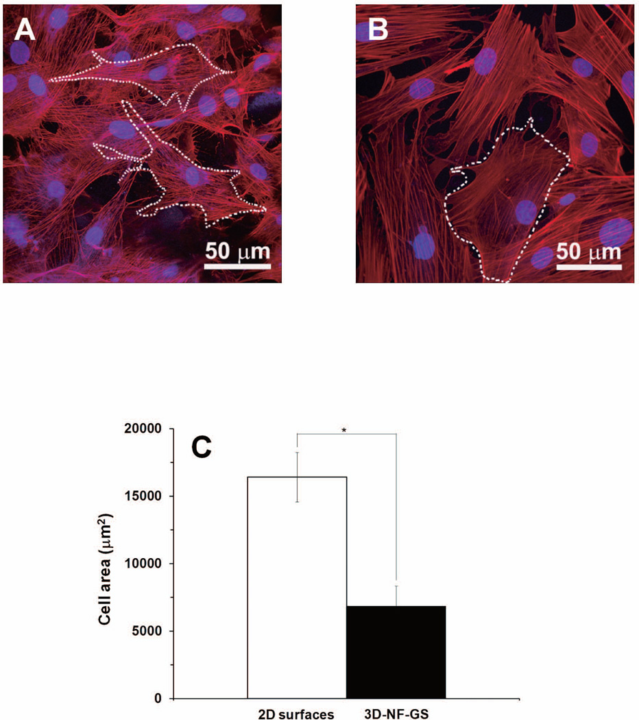
Projected confocal laser scanning microscopy (CLSM) images of osteoblasts on (A) 3D-NF-GS after cultured for 5 days, and (B) 2D gelatin surface. The actin was labeled red and nuclei were blue. (C) Quantification of cell areas on 3D-NF-GS and 2D substrate (*denotes significant difference, p<0.001).

