Figure 1.

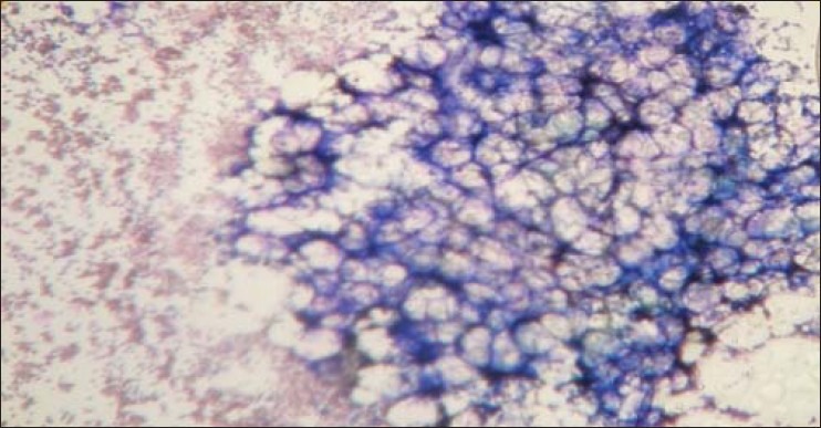
Hypocellular marrow with fat space (aspiration 10X, Leishman stain)Microscopically the aspiration material showed a small nodular area (“hot spots”) consisting of all types of marrow cell, which in turn was surrounded by large fatty spaces

Hypocellular marrow with fat space (aspiration 10X, Leishman stain)Microscopically the aspiration material showed a small nodular area (“hot spots”) consisting of all types of marrow cell, which in turn was surrounded by large fatty spaces