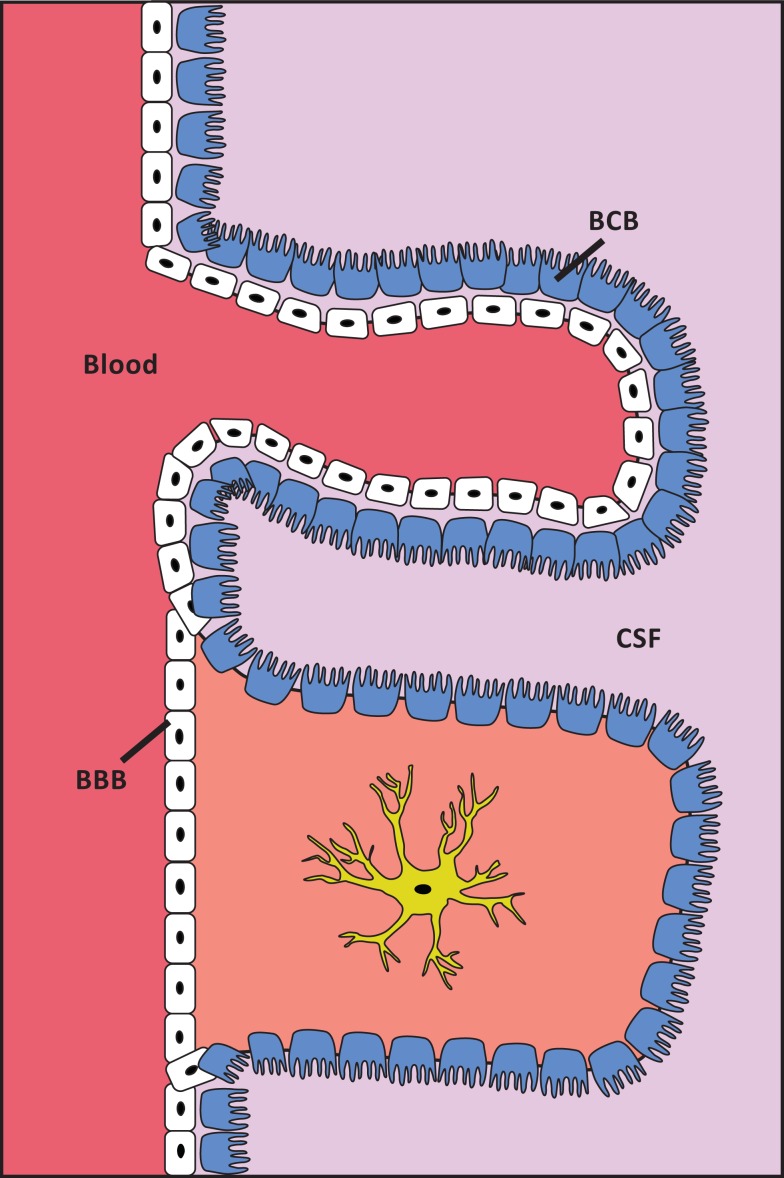
Figure 1.
Schematic presentation of the localization of the BBB and the BCB. White cells are capillary endothelial cells. Blue cells are ependymal cells. The structure of cell layers in the choroid plexus/BCB is shown in the top of the figure. The structure of cell layers elsewhere in the brain/BBB is shown in the lower part of the figure. The capillary endothelial cells are tightly bound by tight junctions, except at the choroid plexus. In the choroid plexus the ependymal cells are, in contrast to elsewhere in the brain, tightly bound by tight junctions.

