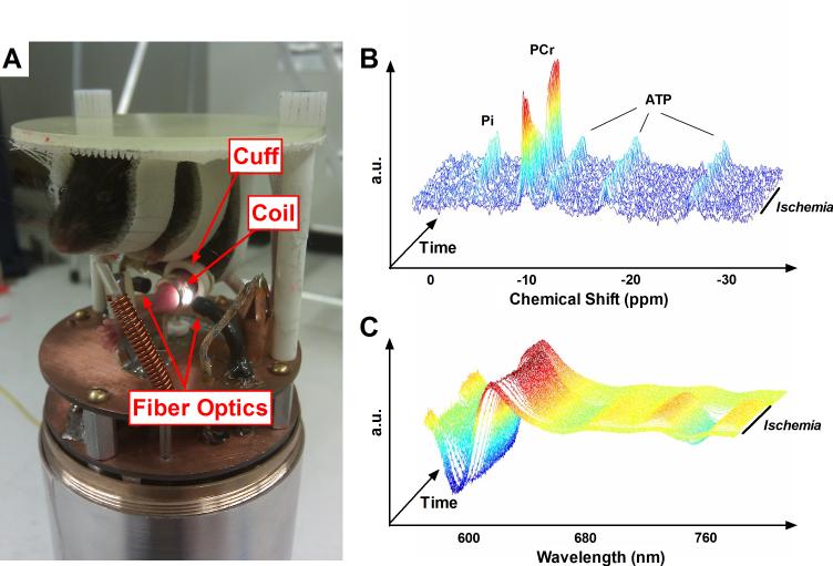
Fig. 1. Multi-modal in vivo spectroscopy.
(A) Photograph of the combined MR and optical spectroscopy probe for use with the Bruker 14T spectrometer. An anesthetized mouse is suspended above spectroscopy hardware, with its shaved distal hindlimb positioned in the center of an MR coil tunable to both 1H and 31P, flanked on either side by fiber optic bundles. An inflatable ischemia cuff surrounds the hindlimb proximal to the MR coil. Unlabeled components visible in the picture include a 100% O2 delivery line, thermocouple, and warm air delivery port. (B) Representative 31P MR spectra (after exponential multiplication, Fourier transformation, and summing 5 consecutive spectra), stacked through the course of a dynamic in vivo experiment. During ischemia, PCr is broken down to maintain constant ATP as the resting energy demands of the cell are met and Pi undergoes a stoichiometric increase. (C) Representative NIR optical spectra (after taking second-derivative) during a dynamic in vivo experiment. As Hb and Mb deoxygenate during ischemia, a pronounced rightward shift is evident between ~580 and 660nm and a dramatic sinusoidal feature becomes prominent at ~760nm.

