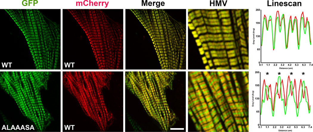
Fig. 4.
The ALAAASA mutant is incorporated into sarcomeres but accumulates in the bare zone. Neonatal rat ventricular myocytes were co-transfected with GFP- and mCherry-WT myosin rods (Top Panel), or with GFP-mutant myosin (ALAAASA) and mCherry-WT myosin rods (Bottom Panel). Representative images from the mCherry and GFP channels are shown together with the merge. HMV: high magnification view of merged images. Linescan: analysis of the relative intensity of wt-mCherry myosin and GFP-myosin mutant along four sarcomeres. Asterisks indicate accumulation of the ALAAASA mutant in the bare zone. The bar represents 10 µm.

