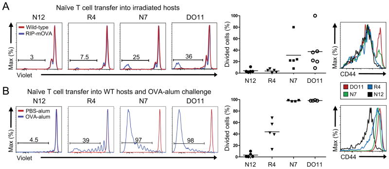
Figure 6. High affinity TCR recognition of peripheral self-antigen is required to elicit peripheral naive T cell responses.
(A) Peripheral T cells responses to RIP-mOVA. Naive peripheral Foxp3−CD4+ T cells were intravenously transferred into sublethally irradiated RIP-mOVA recipients. Representative flow cytometry plots are shown of the transferred splenic T cells after 14 days to determine proliferation via dilution of Cell-Trace Violet dye (left). Frequencies of proliferated cells are summarized for each TCR (middle), and CD44 expression is shown (right). Each dot represents data from a single recipient, with 2 independent experiments per TCR. (B) Peripheral T cell responses to abundant antigen. Naive peripheral Foxp3−CD4+ T cells were intravenously transferred into normal WT recipients immunized with OVA protein-Alum, and proliferation of the transferred T cells in the spleen was analyzed after 7 days. Representative flow cytometric plots of proliferation are shown on the left and summarized in the middle graph. Each dot represents an individual recipient, with 2–3 independent experiments per TCR. Representative CD44 expression is shown on the right.

