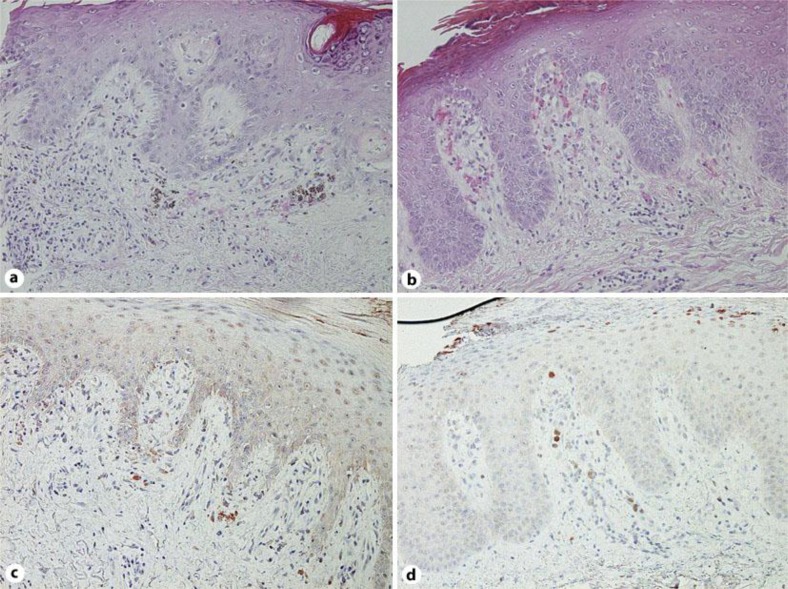
Fig. 2.
Hydropic degeneration of the basal cell layer with band-like infiltration of leukocytes and extravasation of erythrocytes (a). Markedly elongated rete ridges, parakeratosis with neutrophils and dilated tortuous vessels in the dermal papillae (b). Immunohistochemistry for IL-27; paraffin-embedded tissue samples from patients with SLE (c) and psoriasis (d) were deparaffinized and stained using anti-IL-27 Ab (a–d). Original magnification ×200.

