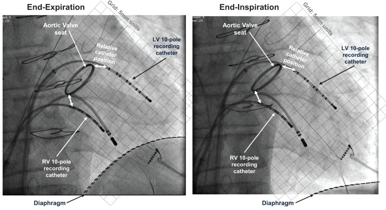
Figure 1.
Electrical recording methods. Fluoroscopic images at end-expiration and end-inspiration of respiratory cycle, both at end-diastole. 10-pole electrode catheters are located in the left and right ventricles, with additional pacing catheter at right ventricular apex. This patient (not included within the population of this study) had a replacement aortic valve with radiopaque aortic ring; this and the sternal wires provide position markers by which to quantify relative positions.

