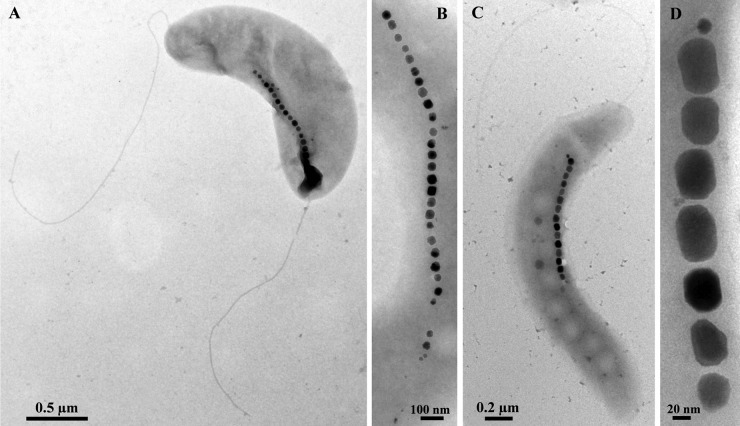
Fig 2.
TEM images of cells of the newly isolated MTB that are slightly different in morphology from Magnetospirillum species. (A) A cell of strain UT-2 negatively stained with 1% uranyl acetate. Note the more vibrioid morphology. (B) Magnetosome chain of strain UT-4 showing cuboctahedral crystals. (C) Cell of strain LM-1 negatively stained with 1% uranyl acetate. Note the presence of a single polar flagellum. (D) High-magnification TEM image of a chain of elongated prismatic crystals of magnetite in a cell of strain LM-1.

